Agricultural and Biological Research
RNI # 24/103/2012-R1
Research - (2023) Volume 39, Issue 6
Algae are the aquatic phototrophic organisms that play a major role in ecosystem. They are the primary producers and have lot of other potential applications. Algae dwells in marine, freshwater, salt water or in any extreme conditions. The goal of this study was to analyses the Morphological and anatomical features of a marine macroalgae Codium collected from Pattukottai. To study the potential activities of macroalgae, phytochemical studies were done. Ethanolic extract of Codium was investigated for the phytochemicals present in the algae. The presence of many types of chemical compounds and particularly high quantity of Vitamin E was present in the macroalgae Codium. Vitamin E is abundant in food and has much beneficial activities on skin care. As a result, the presence of Vitamin E in algae shows that, it can be used on commercial aspects. Codium is considered as edible algae because of the presence of enormous amount of Vitamin E.
Haberler
Haberler
Haberler
Haberler
Haberler
Haberler
Haberler
Haberler
Haberler
Haberler
Haberler
Haberler
Haberler
Haberler
Haberler
Haberler
Haberler
Haberler
Haberler
Haberler
Haberler
Haberler
Haberler
Haberler
Haberler
Haberler
Haberler
Haberler
Haberler
Haberler
Haberler
Haberler
Haberler
Haberler
Haberler
Haberler
Haberler
Haberler
Haberler
Haberler
Haberler
Haberler
Haberler
Haberler
Haberler
Haberler
Haberler
Haberler
Haberler
Macroalgae; Codium; Phytochemical; Vitamin-E
Macroalgae are the primary producers at the base of the marine food web. They feed a variety of communities of herbivorous creatures, including both invertebrates and vertebrates like herbivorous fish, and ultimately act as a safe haven for them from carnivorous predators. To combat this herbivore, which can be aggressive at times, many macroalgae have developed stronger defense systems. Algae are one of the main targets for sustainable biofuel generation and CO2 consumption due to their photosynthetic capabilities. Algae that provide us with our food, fuel for our cars, and the air we breathe are source of active pharmaceuticals that can be utilized to treat malignancies, viruses and drug-resistant bacterial strains [1].
Based on field survey, distinct locations in the Gulf of Mannar are home to four species of Chlorophyta, nine species of Phaeophyta, and nine species of Rhodophyta. Using accepted techniques, 22 different seaweed species were identified and described. The most common classes of algae in nature are Chlorophyceae, Phaeophyceae, and Rhodophyceae, with 900, 1500, 4000 species each [2]. Seaweeds are abundant, have a large biomass potential, and are used in many ways, including as food. Products made from macroalgae are healthy and protective against a number of ailments. The usage of seaweeds as food, gels, and/or emulsifiers in many industries is in addition to their use as animal feed and fertilizer for agriculture. Comparing macroalgae to higher plants reveals fascinating physiological characteristics [3]. In total, 57 taxa from the southern districts of Tamil Nadu-Kanyakumari, Tirunelveli, Tuticorin, and Ramanathapuram-representing Chlorophyta (18 taxa), Ochrophyta (14 taxa), and Rhodophyta (25 taxa) were documented. In terms of species, more algae were found in Idinthakarai (84.21%). The distribution of Gracilaria corticata is 10 locations, or 52.63%. Maximum (59%) and least (1%), respectively, species were found in Idinthakarai-Tirunelveli district and Keelvaipaar-Tuticorin, all of which are located on the Bay of Bengal. The Bay of Bengal coasts had the highest concentration of algae (67.7%), followed by those around the Indian Ocean (25%) Arabian Sea shores (8%) [4].
Sample collection and authentication
The Macroalgae Codium along with other two species were collected from Pattukottai, coastal region. The region is Velivayal, Athiramapattanam post, near Palk Bay. The algal species were collected in three sites at Latitude and longitude of 10.287 23N 79.34248E, 10.28715N 79.34168E, 10.28701N 79.34077E. The collected macroalgae are washed thoroughly with seawater and then washed with distilled water to remove the debris and salt present. Macroalgae species are dried and kept as an herbarium for further identification of species.
Chemicals required
Anatomical studies of Macroalgae Codium are done under the laboratory condition. Saffranine is used for the staining of transverse section of Codium. The stained sections were mounted in the glass slide with the help of glycerine. Mounted specimen in the slide was observed in the Light microscope at different magnifications 4x, 10x, and 40x. For further phytochemical studies the Codium sample was prepared with the help of Ethanol.
Sample preparation
The Codium sample was prepared with ethanol for screening the materials present in the species by Gas chromatography-Mass spectrometry. For that, dried sample of Codium was prepared from the fresh algal specimen of weight 13.084 g. The dried Codium sample was added to 100 ml of Ethanol. The prepared Ethanol sample were filtered with the help of Whatman no1 filter paper and the filtrate were transferred to small vial. This Ethanol sample is used for the screening of chemical compounds present in the sample through Gas chromatography-Mass spectrometry.
GC-MS analysis of ethanolic algae extract
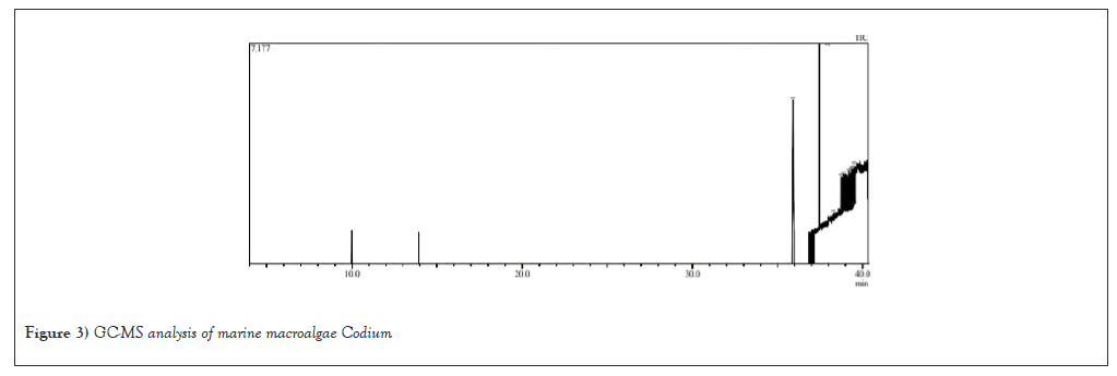
A Ethanolic extract of the marine Macroalgae Codium was analysed by GC-MS using a VF-5ms fused silica capillary column (Model; QP 2010 Plus, Shimadzu, Tokyo, Japan). It was intended for the column oven's temperature to increase from 50°C to 280°C. At 250°C, the injector was set. Helium is the carrier gas, flowing at a rate of 1.20 ml/min. A Hamilton syringe was used to manually inject an ethanolic extract of Codium into the GC-MS during a split injection operation for total ion chromatographic analysis. It took 40 minutes to finish the GC-MS. RTHE elative percentage of each extract constituent were presented as a percentage using the peak area normalization. By comparing retention indices and mass spectral patterns with sources from the Fatty Acid Methyl Ester Library version 1.0 and the Willey Registry of Mass Spectral Data (Wiley 8), the bioactive components in ethanol were found (FAME library). Retention time is time between the initial injection and the Time of Recovery (RT). To quantify the components in the injected sample, the signal strength was measured on the Y-axis while the RT was displayed on the X-axis. The mass/charge (M/Z) ratio was calibrated using the acquired graph, also referred to as the mass spectrum graph, which is a fingerprint of the molecular structure. By comparing retention indices and spectroscopic fragmentation patterns to those found in computer libraries and published literature, it was possible to identify the components of the extracts.
Morphology of Codium: Codium, Chlorophyceae comprise of following characters;
Division: Chlorophyta
Class: Ulvophyceae
Order: Bryopsidales
Family: Codiaceae
Genus: Codium
External features
Codium is dark green algae that can grow to a height of 10-40 cm. Its segments are frequently branched and cylindrical, measuring 0.5 to 1.0 cm in diameter and the branches can be as thick as pencils. The segments resemble thick, green fingers. A big, sponge-like cushion of tissue serves as its holdfast.
Anatomy of codium
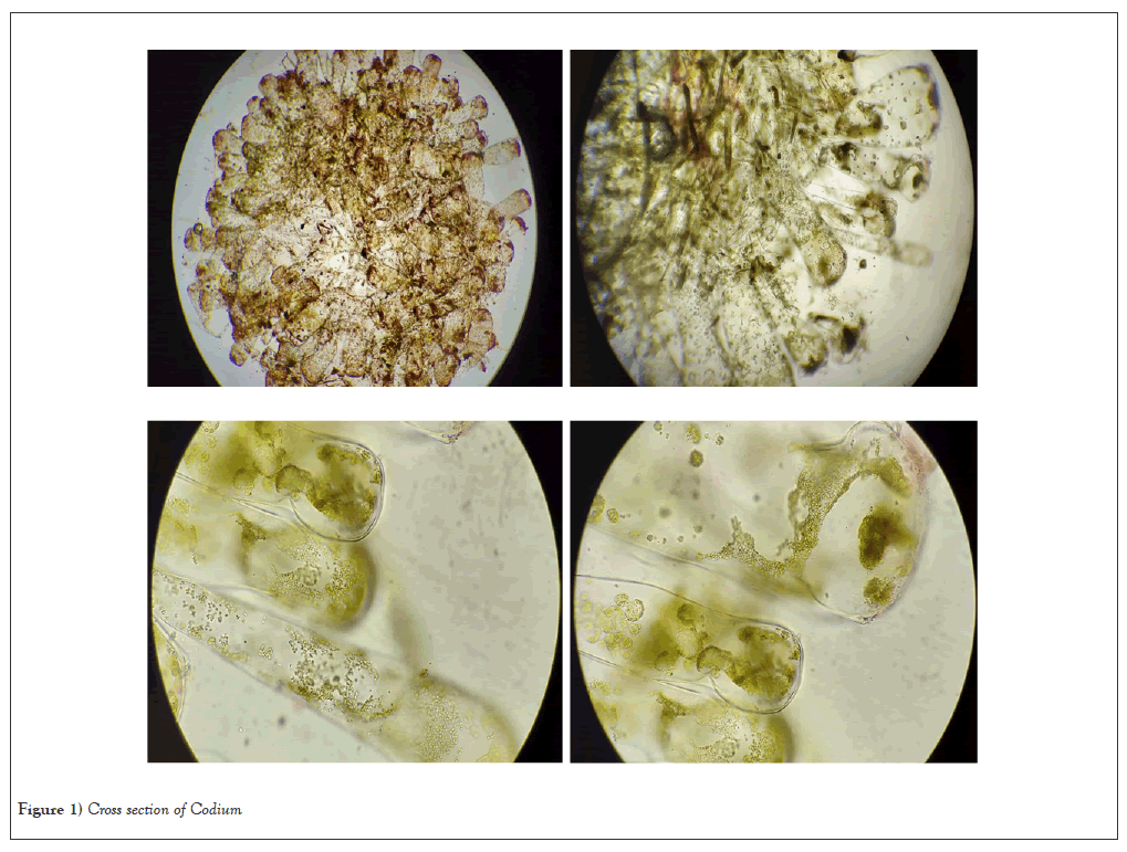
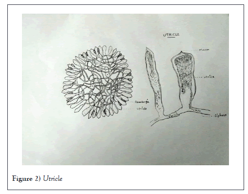
Anatomy of Codium thallus is composed of the single, large, branching tubular cell with many nuclei; branches are sometimes referred to as siphons. The thallus's center is made up of entangled web of siphons, in contrast to the closely packed, utricle-forming cortex that surrounds it. The content, size, and form of utricles might vary. Moreover, they could have gametangia or side hairs. Medullary filaments are slender outgrowths from the base of each utricle that range in diameter from 26 to 46 µm, sharply defining the boundary between the utricle and filament. Utricles range in size from 130 to 400 µm in width and 400 to 1000 µm in length. Many are conical in shape with thicker apex walls. Typically, these utricles have short bulbous shape. Hairs attached to darkened swollen tip. Gametangia is elongate-ellipsoidal, sometimes ovate, 80-130 µm diameter 260-330 µm long, born on short bud distinct pedicle at or just above middle of utricle, 1-3 per utricle, extending to apex of the utricle. The utricles, a palisade-like layer of green vesicles that make up the interior structure, are interwoven densely in the colorless medulla. Organelles are restricted to a layer of cytoplasm connected to the cell wall, of which mannan is a crucial component, including several nuclei and disc-shaped chloroplasts. Chloroplasts do not include pyrenoids; siphaxanthin and siphonanthin are carotenoid pigments. The centripetal deposition of wall material results in incomplete septa (plugs). Utricles are primarily created by the growth of medullary siphon sympodial branches, with secondary production occurring by budding or the generation of new medullary siphons that produce utricles from base of pre-existing utricles. The apical wall of mature utricles is frequently thicker and frequently adorned with a pattern particular to a given species. They are clavate or cylindrical. Rhizoid siphons are another product of the basal part of utricles; they burrow into the medulla. Any of colorless hairs has basal clog produced by utricles near the hair's apex. Although being caducous, the plug leaves a visible scar. Utricles feature a basal plug above a short pedicel, are fusiform to oval in shape, and generate gametangia laterally. The gametangia's contents split into biflagellate gametes during meiosis. Gametes came out of the apical breach as a gelatinous mass. Although female gametes are larger and contain a greater number of chloroplasts than male gametes, male and female gametes can sometimes develop on the same thallus (monoecious or dioecious). The zygote develops upright, protracted vesicles after becoming an amorphous, prostrate vesicle. The major utricle-producing siphons that emerge from these vesicles eventually unite to form a multiaxial thallus. Through parthenogenesis, fragmenting or chopping off modified aborted gametia, or other asexual reproductive techniques (Figures 1 and 2).

Figure 1: Cross section of Codium.

Figure 2: Utricle.
Reproduction
In Codium, only gametophyte generation takes place. The separate chambers in which the sperm and eggs are housed are called gametangia, and they protrude from the utricles. The majority of the dioecious Codium species have male and female gametangia on separate plants. Both male and female gametes have two flagella, but the female gametes are larger.
Gametogenesis
Male gametogenesis: On the sidewalls of the utricules, gametangia developed. Gametangia first displayed vegetative interphase nuclei and the chloroplasts that already had a mature structure with a limited number of thylakoid lamellae and single starch granule present. Then there were divisions known as mitosis, which led to an increase in gametic nuclei. The gametangial mitosis shared characteristics with other Bryopsidophyceae's vegetative mitosis. The union of tiny vesicles with a big mitochondrion and chloroplasts clearly separated uninucleate regions of protoplasm. The basal-flagellar apparatus eventually evolved once the gamete's ultimate shape was defined. Via an apical aperture, mature gametes were released and submerged in a mucilaginous substance. They had an elongated-ovoid form, two flagella, a hyaline front section, and rear portion containing the two or three yellow-green chloroplasts. The anterior nucleus was densely packed with chromatin but lacked a nucleolus. At the front of the gamete was a sizable mitochondrion that had an "inverted-V" form. Lamellae in chloroplasts were underdeveloped, and the starch granules were big.
Female gametogenesis: The protoplasm was homogeneous early in gametogenesis but later became fragmented as the number of fusiform nuclei and chloroplasts increased. Other Bryopsidophyceae shared a number of traits with the nuclear divisions. They were somewhat acentric and open. Indicating meiosis were parallel electron-dense lines in number of prophase nuclei that resembled synaptonemal complexes. During metaphase, the nuclear envelope showed polar fenestrae that gave rise to the spindle. Chromosomal kinetochores were present, but spindle microtubule-nucleating material was not. On the pyriform, hyaline anterior end of the mature female gametes, there were two flagella. The spherical nucleus of mature gametes was ringed by a mitochondrion and many discoid chloroplasts. Female gametes germinate parthenogenetically in both gametangia and in culture, losing their flagella, rounding, cell lengthening proliferating chloroplasts with well-established thylakoid systems; vacuolizing and developing the fibrillar cell wall (Table 1 and Figure 3).
| S. No | Ret. Time | Ret. Index | SI | Area% | Compound name | Molecular formula | Molecular Weight |
|---|---|---|---|---|---|---|---|
| 1 | 35.903 | 3149 | 89 | 38.23 | Vitamin E | C29H50O2 | 430 |
| 2 | 37.462 | 0 | 79 | 19.49 | 1,2-Benzenedicarboxylic acid, Dioctyl ester | C24H38O4 | 390 |
| 3 | 38.304 | 1767 | 100 | 0.38 | Indolizine, 2-(4-Methylphenyl) | C15H13N | 207 |
| 4 | 38.73 | 0 | 94 | 3.69 | 3,4-Dihydro-4-(1,3-Dioxolan-2-YL)-5,7-Dimethoxy-1(2H)-Benzopyran-2-One | C14H16O6 | 280 |
| 5 | 38.885 | 942 | 100 | 6.91 | (SS)- or (RR)-2,3-hexanediol | C6H14O2 | 118 |
| 6 | 39.186 | 942 | 95 | 9.68 | (SS)- or (RR)-2,3-hexanediol | C6H14O2 | 118 |
| 7 | 39.29 | 0 | 93 | 1.3 | 2((Trimethylsilyl)Ethynyl) Heptamethyltrisilane | C12H30Si4 | 286 |
| 8 | 39.365 | 0 | 96 | 9.04 | 2((Trimethylsilyl)ethynyl) Heptamethyltrisilane | C12H30Si4 | 286 |
| 9 | 39.38 | 0 | 93 | 1.37 | 2((Trimethylsilyl)ethynyl) Heptamethyltrisilane | C12H30Si4 | 286 |
| 10 | 39.505 | 942 | 97 | 9.9 | (SS)- or (RR)-2,3-hexanediol | C6H14O2 | 118 |
Table 1: Phytochemicals after gas chromatography mass spectrometry analysis.

Figure 3: GC-MS analysis of marine macroalgae Codium.
Phytochemicals in codium
Vitamin-E: A fat-soluble nutrient called vitamin E can be taken as a dietary supplement or added to various meals. Vitamin E functions as an antioxidant in the body, helping to shield cells from damage brought on by free radicals. When our body transforms the food we eat into energy, chemicals called free radicals are created. Vitamin E supports healthy skin, eyes, and the body's natural defenses against disease and infection. Supplemental vitamin E may be beneficial for those with specific skin conditions like eczema. Vitamin E supplements may be helpful for those with certain skin disorders such as eczema. It is an essential nutrient that plays a crucial role in several biological functions, including
Antioxidant activity: Vitamin E is a potent antioxidant that protects cells from damage caused by free radicals. Free radicals are highly reactive molecules that can damage cells and contribute to the development of chronic diseases, including cancer, heart disease, and Alzheimer's disease.
Immune system support: Vitamin E helps support the immune system by enhancing the activity of immune cells, such as T cells and B cells. It also reduces inflammation, which is a key factor in many immune-related diseases.
Skin health: Vitamin E is important for maintaining healthy skin. It helps to protect the skin from UV damage, reduces inflammation, and promotes wound healing.
Cardiovascular health: Vitamin E has been shown to help reduce the risk of heart disease by preventing the oxidation of LDL cholesterol, which can lead to the formation of plaque in the arteries.
Brain health: Vitamin E may help protect against cognitive decline and Alzheimer's disease by reducing oxidative stress and inflammation in the brain.
Anti-cancer properties: Vitamin E has been shown to have anti-cancer properties, particularly in the prevention of colon, breast, and prostate cancers. It may help prevent the growth of cancer cells by reducing oxidative stress and inflammation. Overall, Vitamin E is an important nutrient that plays a critical role in maintaining good health and preventing chronic diseases [5].
1,2-Benzenedicarboxilic acid, Diocytl ester
Phthalic acid is another name for benzoenedicarboxylic acid. A class of lipophilic compounds known as Phthalic Acid Esters (PAEs) are frequently employed as plasticizers and additives to increase the mechanical extensibility and flexibility of a variety of products. In addition, PAEs are regularly found in sources from plants and microorganisms, raising the idea that they are produced naturally. In addition to being discovered in a wide range of diverse plant species' organic solvent extracts, essential oils and root exudates, PAEs have also been isolated and purified from several bacteria, fungi, and algae. At least a few types of algae are capable of biosynthesising PAEs. Allelopathic, antibacterial, insecticidal, and other biological activities of PAEs have been documented. These biological activities may increase the competitiveness of algae, plants, microorganisms to better withstand biotic and abiotic stress. Due to their widespread synthesis and use, these findings show that PAEs shouldn't be only regarded as "human-made pollutants". On the other hand, manufactured PAEs that enter the ecosystem may affect the metabolism of some plant, algal, and microbial populations. Hence, additional research is necessary to clarify the pertinent mechanisms and ecological effects [6].
Indolizine, 2-(4-methylphenyl)
The indolizine family of heterocyclic compounds includes the synthetic chemical indolizine, 2-(4-methylphenyl). It has been investigated for potential biological activity and has produced encouraging outcomes in a number of experiments. Here are a few of 2-(4-methylphenyl) indolizine's biological activities that have been noted:
Anticancer activity: MCF-7, HepG2, and HCT116 are only a few of the cancer cell lines against which Indolizine, 2-(4-methylphenyl), has demonstrated substantial cytotoxic action.
Anti-inflammatory action: By preventing the synthesis of pro-inflammatory cytokines including TNF-alpha, IL-6, and IL-1beta, Indolizine, 2-(4-methylphenyl), has demonstrated a possible anti-inflammatory effect.
Antimicrobial activity: Indolizine, 2-(4-methylphenyl) has shown significant antimicrobial activity against various bacterial strains such as Staphylococcus aureus, Escherichia coli, and Pseudomonas aeruginosa.
Antioxidant activity: Indolizine, 2-(4-methylphenyl) has shown significant antioxidant activity by scavenging free radicals and inhibiting lipid peroxidation.
Antiviral activity: Indolizine, 2-(4-methylphenyl) has shown promising antiviral activity against the Herpes Simplex Virus type 1 (HSV-1).
It is important to note that the biological activities of Indolizine, 2-(4-methylphenyl) have mainly been studied in vitro and further studies are required to evaluate its potential in vivo efficacy and safety [7].
3,4-Dihydro-4-(1,3- dioxolan-2yl)-5,7--dimethoxy-1(2h)-benzopyran-2-one
3,4-dihydro-4-(1,3-dioxolan-2yl) -5,7-dimethoxy-1(2h) Dihydrocoumarin and coumarin-3,4-dihydro-2h-1-benzopyran-2-one are other names for benzopyran-2-one. It is a naturally occurring substance that is present in many plants, including tonka beans, cinnamon, and sweet clover. It's utilized in flavorings, cosmetics, and fragrances because of its sweet, vanilla-like scent. For many years, coumarin has been utilized as a food flavoring, especially in baked goods like cakes, pastries, and bread. Because of its potential for toxicity at high levels, its usage has been limited or outright prohibited in a number of nations. Except for some traditional and seasonal foods, the European Union has established a maximum limit of 1 mg/kg for coumarin in foodstuffs. The Food and Drug Administration (FDA) in the United States has not set any precise limits on the amount of coumarin that may be used in food, but it has listed coumarin as a flavoring ingredient that is Generally Recognized as Safe when used in line with good manufacturing practice. Although modest coumarin use is generally regarded as safe, excessive doses have been associated with liver damage, especially in people who already have liver issues. Because of this, it's crucial to be aware of potential coumarin sources in food and use it sparingly [8].
(SS)-or (RR)-2,3-hexanediol
A type of diol having the chemical formula C6H14O2 is hexanediol. It can be used in a broad variety of processes, such as the synthesis of different compounds and as a plasticizer, humectant, solvent, and plasticizer. Hexanediol has demonstrated biological actions that have antibacterial qualities. A number of bacterial species, including Staphylococcus aureus, Escherichia coli, and Pseudomonas aeruginosa, have been discovered to be particularly susceptible to it. Also employed in dermatology, hexanediol has been found to have moisturizing and emollient qualities. To keep the skin moisturized and supple, it can be found in many skincare products, like lotions and moisturizers. Hexanediol has also been researched for its possible use as a transdermal medication delivery penetration enhancer. According to studies, it can enhance the skin absorption of several medications, enabling a more effective and efficient administration of these substances.
Anatomical studies were done under laboratory condition. Phytochemical work done on macroalgae Codium from that we came to know the presence of essential chemical compounds. GC-MS result shows that vitamin E content (36%) high in Codium. Vitamin E which mostly present in many plant-based foods which are edible. Vitamin E helps in maintain healthy skin and eyes, and strengthen the body’s natural defense against illness and infection. It also has Antimicrobial and Antioxidant properties. So that Codium is considered as a macroalgae which can be commercially used. Algae growing in stressed condition have enhanced phytocompounds and more productivity. The hydrocarbon storage as fatty acids and pigmented compounds helps to thrive the algae in the stressed condition [9]. The tocopherol constituent in the Codium is vast higher than the remaining macroalgae like Dictyota, Gracillaria from the same collection site [10-36].
Based on the result of this study, the phytochemical composition of macroalgae Codium has been determined. Ethanolic extract of Codium contain important and active phytochemical compounds which are used mostly in commercial and therapeutic aspects. Each compound which presents has biological properties. Their specific role has been investigated in this study. The phytochemicals which present are Vitamin-E-38%, 1,2-Benzenedicarboxylic acid, Dioctyl Ester-19%, Indolizine, 2-(4-Methylphenyl)-0.38%, (SS)- or (RR)-2,3-hexanediol-9%, 3,4-dihydro-4-(1,3-dioxolan-2yl) -5,7-dimethoxy-1(2h)-benzopyran-2-one-3%. It has been determined that most active compound in Codium is Vitamin-E. 1,2-Benzenedicarboxylic acid, Dioctyl Ester which has antimicrobial properties. Indolizine,2-(4-Methylphenyl) has antimicrobial, anticancer, antiviral, and antioxidant activities. (SS)- or (RR)-2,3-hexanediol has been used in skin care products. 3,4-dihydro-4-(1,3-dioxolan-2yl)-5,7-dimethoxy-1(2h)-benzopyran-2-one has aromatic compounds, so that it is used to increase flavors, and mostly present in cinnamon, sweet clover. Vitamin-E also called as alpha-tocopherol which are mostly present in edible plant-based products. Vitamin-E is an antioxidant. It helps to keep strong immune system. Therefore, in this study, presence of Vitamin-E in the extract can be considered to be involved in food, medicine and cosmetics. And also, Vitamin-E involves protecting skin from solar radiation. So, it also been widely used in skin care products and cosmetics. The presence of phytochemicals in the extract confirms that, this Macroalgae can be used commercially in food, medicine and cosmetics. In addition, the presence of higher percentage of Vitamin-E proves that Codium can also be taken as dietary supplement.
[Crossref] [Google Scholar] [PubMed]
[Crossref] [Google Scholar] [PubMed]
[Crossref] [Google Scholar] [PubMed]
[Google Scholar] [PubMed]
[Crossref] [Google Scholar] [PubMed]
[Crossref] [Google Scholar] [PubMed]
[Crossref] [Google Scholar] [PubMed]
[Crossref] [Google Scholar] [PubMed]
[Crossref] [Google Scholar] [PubMed]
Citation: Deepa KP, Thivyadharshini M, Gideon AV, et al. Anatomical and phytochemical properties of Codium, a marine macroalga. AGBIR.2023;39(6):682-687.
Received: 10-Oct-2023, Manuscript No. AGBIR-23-117357; , Pre QC No. AGBIR-23-117357 (PQ); Editor assigned: 13-Oct-2023, Pre QC No. AGBIR-23-117357 (PQ); Reviewed: 30-Oct-2023, QC No. AGBIR-23-117357; Revised: 08-Nov-2023, Manuscript No. AGBIR-23-117357 (R); Published: 15-Nov-2023, DOI: 10.35248/0970-1907.23.39.682-687
Copyright: This open-access article is distributed under the terms of the Creative Commons Attribution Non-Commercial License (CC BY-NC) (http:// creativecommons.org/licenses/by-nc/4.0/), which permits reuse, distribution and reproduction of the article, provided that the original work is properly cited and the reuse is restricted to noncommercial purposes. For commercial reuse, contact reprints@pulsus.com This is an open access article distributed under the terms of the Creative Commons Attribution License, which permits unrestricted use, distribution, and reproduction in any medium, provided the original work is properly cited.
